


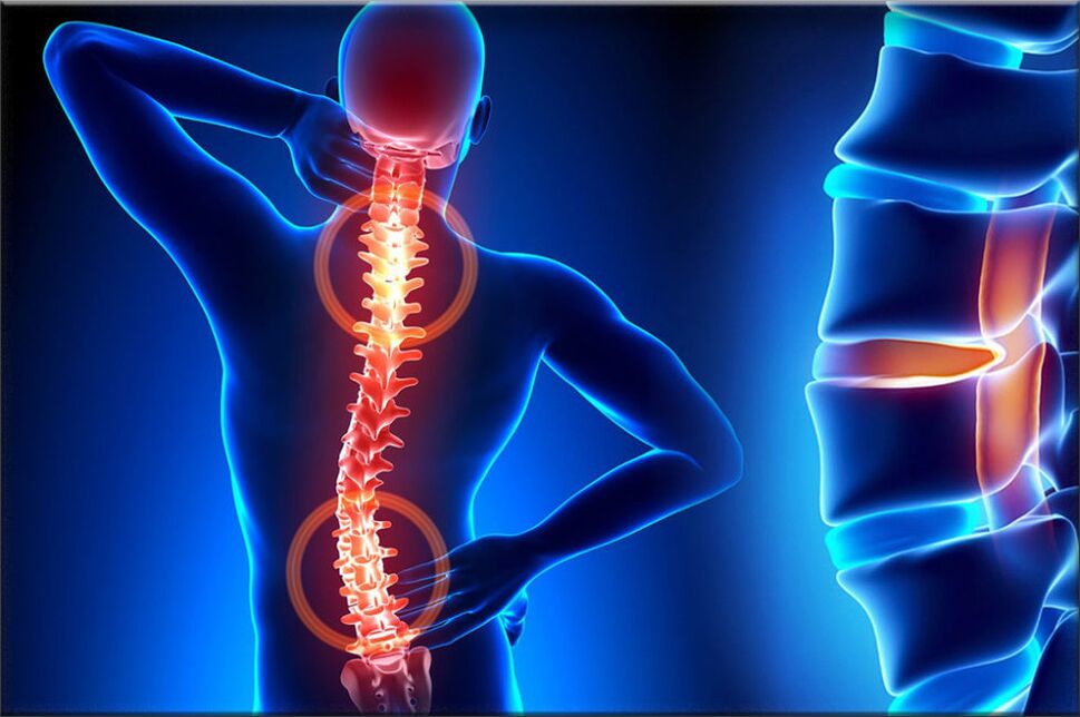
Osteochondrosis is one of the most common diseases of the musculoskeletal system. It is a comprehensive result of certain dystrophic changes in spinal cartilage. During this pathological process, the intervertebral discs of the spine are often affected. The structures are intervertebral cartilage discs that provide flexibility and also allow the person's spine to move, that is, they provide movement.
In the case of osteochondrosis, many processes occur that cause the intervertebral disc to degenerate. As a result, the intervertebral disc begins to lose elasticity and reduce flexibility, and at this time the intervertebral disc itself becomes very flat. The distance between the two intervertebral discs is reduced, which simultaneously compresses nerve endings and blood vessels and causes severe pain. The compressed area of the nerve node begins to swell, causing the pain to worsen or even worse.
In the development of osteochondrosis, muscle structure and most organs of the body often participate in this pathological process. This is due to the fact that blood circulation and the activities of muscles and organs are disturbed in the process of maximizing the violation of neurovascular bundles. For example, the most common osteochondrosis is cervical osteochondrosis, which is accompanied by head and back pain, nausea, dizziness, visual impairment, and tinnitus. This disease has become quite "young": a century ago, osteochondrosis was a disease of elderly patients, and now young people are also vulnerable to this disease.

The most vulnerable people are those who severely impair the body's metabolism and hormone levels, as well as those who suffer from vascular and venous diseases. This is because these diseases can lead to the destruction of disk oxygenation. If timely and appropriate measures are not taken to heal, the edge of the compressed intervertebral disc will be compressed and will exceed the limits of the spine anatomically, thereby destroying the neurovascular bundle.
Therefore, the patient is at risk of herniated disc. The main cause of osteochondrosis is uneven load distribution in the spine, which leads to changes in the cartilage structure at locations with excessive pressure. The nature of this disease depends on the degree and extent of damage to the affected intervertebral disc. The intervertebral disc changes with age, just like our hair. Severe injuries or fractures to the spine can affect its function. Casual clothing and certain types of vibration can also accelerate the rate of spinal degeneration. In addition, there is evidence that smoking increases the speed of spinal degeneration. Scientists have also discovered connections between family members, highlighting the role of genetics in the speed of change.
The disease can also be triggered by many factors:
- Injuries, bruises;
- Spinal muscular dystrophy;
- Curvature and curvature of the spine;
- weightlifting;
- Stay in one position for a long time;
- Metabolic disease
- Lack of trace elements and vitamins-manganese, magnesium, zinc and vitamins D and F;
- Genetic predisposition
- Body overload
- A sedentary lifestyle;
- Radiation background
- frostbite;
- Congenital malnutrition
- Asymmetrical work of spinal muscles;
- Stress, frustration.
These causes of osteochondrosis are only the hypothesis of scientists and are the direct factors that cause the disease. Science has not yet discovered it. We are only talking about risk factors.
the first lessonDevelopment-characteristic is the early deployment of the nucleus pulposus of the intervertebral disc (the nucleus pulposus of the eccentric disc, located on the dorsal side of the vertebral body).
second stageIt is characterized by the instability of the spinal segment. The pathological substrate is represented by the fiber core of the affected intervertebral disc, which has a degenerative process of shedding and rupture of the posterior longitudinal ligament, thereby forming pathological movement between the vertebrae.
Third periodThe development of the disease-the overall damage of the intervertebral disc, and the appearance of "intervertebral disc herniation"-the dislocation and discharge of the nucleus pulposus fragments outside the intervertebral disc.
If the disease has entered the third stage, the destruction process is already irreversible and may lead to severe disability.
Types of osteochondrosis
The development of osteochondrosis is slow, and the condition is aggravated by spinal injury, exercise, and weightlifting. Clinically depends on the location of the lesion.
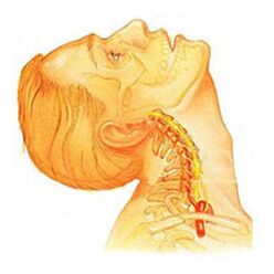
Cervical osteochondrosisLocal and remote symptoms with an advanced form-the roots dominate, that is, it can cause severe radiculopathy. The symptoms of cervical spine osteochondrosis are accompanied by varying degrees of dysfunction, sometimes manifested as a sudden restriction of cervical spine mobility and dysfunction. Headaches can be traction and paroxysmal at the same time by irradiating the inter-shoulder region or the shoulder region. In the acute phase, the patient is diagnosed with an episode of neck pain, which hinders and restricts the movement of the head and neck. In addition to severe discomfort, pain syndrome can also be accompanied by dizziness, insomnia, pain, loss of appetite, depression, and eye and pharynx disorders.
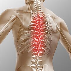
Sternal osteochondrosis. . . The clinical manifestations are caused by the process of local disease and nerve root structure destruction. Sternal osteochondrosis has obvious pain syndrome, which can have chronic or acute back pain, accompanied by chest discomfort and limited muscle contracture, until right speech muscle atrophy. Chest pain can be diffuse, intercostal and neurogenic. Palpation can enhance the axial rotation of the vertebral body. The disease corresponds to the degree of root stimulation from Th11 to Th12, and may manifest as angina pectoris, manifested as liver and gastrointestinal dysfunction. Diseases of the genitourinary system and genital area often occur. Patients pay attention to paresthesias, such as paresthesias, the superficial and deep sensitivity is significantly reduced.
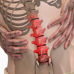
Lumbar osteochondrosis. . . It is characterized by abdominal reflexes and lower limb dysfunction. In the development of neurological diseases, leg muscle weakness and pelvic organ dysfunction may occur. Osteochondrosis is characterized by assessment of damage to the sitting process. The more advanced the stage of lumbar disease, the shorter the time the patient can sit. The lumbar form is characterized by chronic and acute back pain, paraspinal muscle spasm, and myofascial secondary syndrome. The pain will radiate to the hip and posterior bone.
Depending on the locality of the pathological process of osteochondrosis, the disease can cause the patient to violate the surface sensitivity (touch, heat). Changes in reflexes (for example, no Achilles reflex), muscle wasting, dystonia, autonomic disorders (paleness, redness of the skin, nutritional changes in the nails, low temperature of the distal skin), and sphincter dysfunction are also characteristicAnd sexual dysfunction.
Clinical picture
Diagnostic procedureFirst, you must have a complete medical history and physical examination. The doctor asks questions about symptoms and how the disease interferes with the patient's daily activities. In addition, experts are interested in determining postures and activities that emphasize or reduce pain levels.
Then, the doctor examines the patient to check the position and range of movement in the spine to determine the movement that is causing the pain. Skin sensitivity, muscle strength and reflex were all tested in the same way. Based on the medical history and physical examination, the doctor determines which techniques will help.
Radiography is rarely helpful for diagnosis. In the early stages of disease development, the proportion of radiographic images showing abnormalities does not exceed 30%.
However, if the symptoms are severe and the disease has entered the second or third stage, one or more disc defects can be seen in the image. They can be penetrated by osteophytes between the vertebrae and joints.
If other information is needed, magnetic resonance imaging is required. MRI is used to view the soft tissues of the body. This is useful if the core of the tissue absorbs water or there are cracks inside the disc. MRI can show other soft tissue problems, such as spinal nerves.
Records can help with diagnosis. The examination is performed using a contrast agent, which is injected into one or more intervertebral discs. Follow-up observations of radiography provide useful information about the condition of the disc.
The treatment of osteochondrosis depends on the species
Non-surgical treatment of osteochondrosis
Whenever possible, doctors will choose non-surgical treatment. The most important thing in non-surgical treatment is to relieve pain and other discomforts so that patients can restore a comfortable standard of living as much as possible.
Doctors rarely prescribe bed rest for patients with osteochondrosis. Regardless of pain, encourage patients to move naturally. If the symptoms are severe, you can stay in bed for a few days.
When the spine is displaced, an elastic band is sometimes prescribed, and the elastic band should not be worn for more than 2-4 days to avoid back muscle atrophy.
Osteopathy can reduce osteochondrosis.OsteopathIt can not only diagnose the problem area, but also relieve the pain of 1-2 doses, reduce the overall condition of the body and "tighten" the internal organs.
Some medications can be prescribed to the patient to control symptoms and return to normal activities for a long time. If symptoms continue to restrict the patient’s activities, routine doctors may recommend epidural steroid injections.
Steroids are effective anti-inflammatory drugs that help relieve pain and inflammation. Non-steroidal anti-inflammatory injections are injected into the space around the roots of the spine. This area is called the epidural space. Some doctors only inject steroids. However, it is usually combined with other drugs. Basically, steroids are prescribed only when other drugs are ineffective, but osteopathy almost always helps.
In addition, patients often work with physical therapists. After assessing the patient's condition, the therapist will prescribe some exercises to relieve symptoms. This exercise program is designed to improve flexibility and helps train the abdominal and back muscles to exercise with minimal pain.
surgery
People with osteochondrosis usually do not need surgery. In fact, only 1-3% can be operated. Before considering surgery, the surgeon prescribes a non-surgical treatment for at least 3 months, that is, skull ac bone disease as a rehabilitation treatment. If no results are obtained after 3 months of non-surgical treatment, there is only a reason for the surgical procedure.
Basic surgical procedures
Discectomy
This procedure aims to partially or completely remove the intervertebral discs in the lumbar region. Surgeons usually perform surgery through an incision in the waist area. Before removing the herniated disc, it is necessary to remove some flat plates.
Nowadays, surgery has mastered minimally invasive techniques, and only a small incision is required in the lumbar region. Those who support this method claim that it is safe. They also believe that the procedure can prevent scars around nerves and joints and help patients recover faster.
merge
This is an intervention that joins two or more bones together to prevent the ends of bones and joints from wearing away.
recovery
The doctor may advise the patient to see a physical therapist several times a week for 4-6 weeks. In some cases, patients need other help.
Symptoms need to be controlled during the first year of treatment. The therapist will work with you to find postures and movements to relieve pain. Heat, cold, ultrasound and electrical stimulation can be prescribed to relieve pain and muscle spasms. Massage or special forms of soft tissue mobilization can also be used. These procedures can help patients to exercise easily.
Usually, adjusting the treatment method can help restore spinal nerve and muscle sensitivity, reduce pain and improve mobility.
The main purpose of treatment is to teach patients how to operate to prevent problems in the future. Patients are advised to perform a series of exercises to improve flexibility. It will also provide patients with a strategy to help in the event of recurring symptoms.
Everyone should study and consider all types of osteochondrosis to prevent themselves and their loved ones from contracting this disease. After all, it is impossible to treat damaged vertebrae. Treatment is designed to relieve pain symptoms and achieve long-term relief. You also need to remember a simple but effective rule:The best treatment is prevention. . .
Prevent osteochondrosis
Prevention is very simple-it is a healthy diet, regular muscle activity, a warm-up every morning, a healthy and active lifestyle and monthly visits.OsteopathyUsed to correct and eliminate musculoskeletal tension. Following these rules is enough to avoid facing the above-mentioned problems, and avoid terrible symptoms and lifelong treatment.